Exact Nuclei Counts foryour Single Cell Sequencing Workflow
Research Use Only. Not for Diagnostic Procedures.
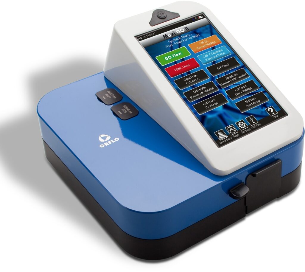
Moxi GO II
Single Cell Sequencing (SCS) workflows can be very expensive, time-consuming, and often require very accurate nuclei counts at low concentrations to ensure your downstream experiments are successful. After all, you can’t perform single cell sequencing studies with doublettes or voids. The Moxi GO II vastly outperforms all existing vision systems by being able to easily detect single nuclei using the volumetric Coulter Principle. The Moxi GO II also excels at low particle concentrations because we count thousands of cells in each test, instead of hundreds, as vision counters do.
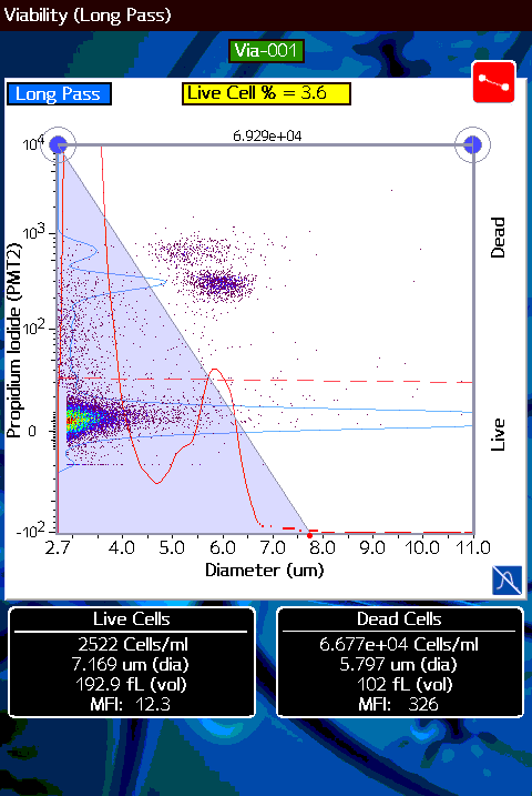
Nuclei isolated from peripheral blood. Distinct nuclei clusters are visible in the upper region of the dot plot. Nuclei are clearly resolved in two dimensions: 1) Coulter-Principle Sizing (x-axis, red histogram overlay shows the clearly-distinguished nuclei size peak) 2) Propidium iodide (Moxi Cyte Viability Kit, MXA055) fluorescence (y-axis, blue histogram overlay shows the fluorescence peak(s)).
SCS Counts you can Trust
Advantages over Vision Systems:
Count, Size, and viability accuracy with 3% typical CVs
Measures particles down to 3μm in (Nuclei, RBC, WBCs, yeast)
Superior Debris Cell Differentiation
Smart Algorithms allow for auto-gating of Viability Tests for Consistent Results