


 |
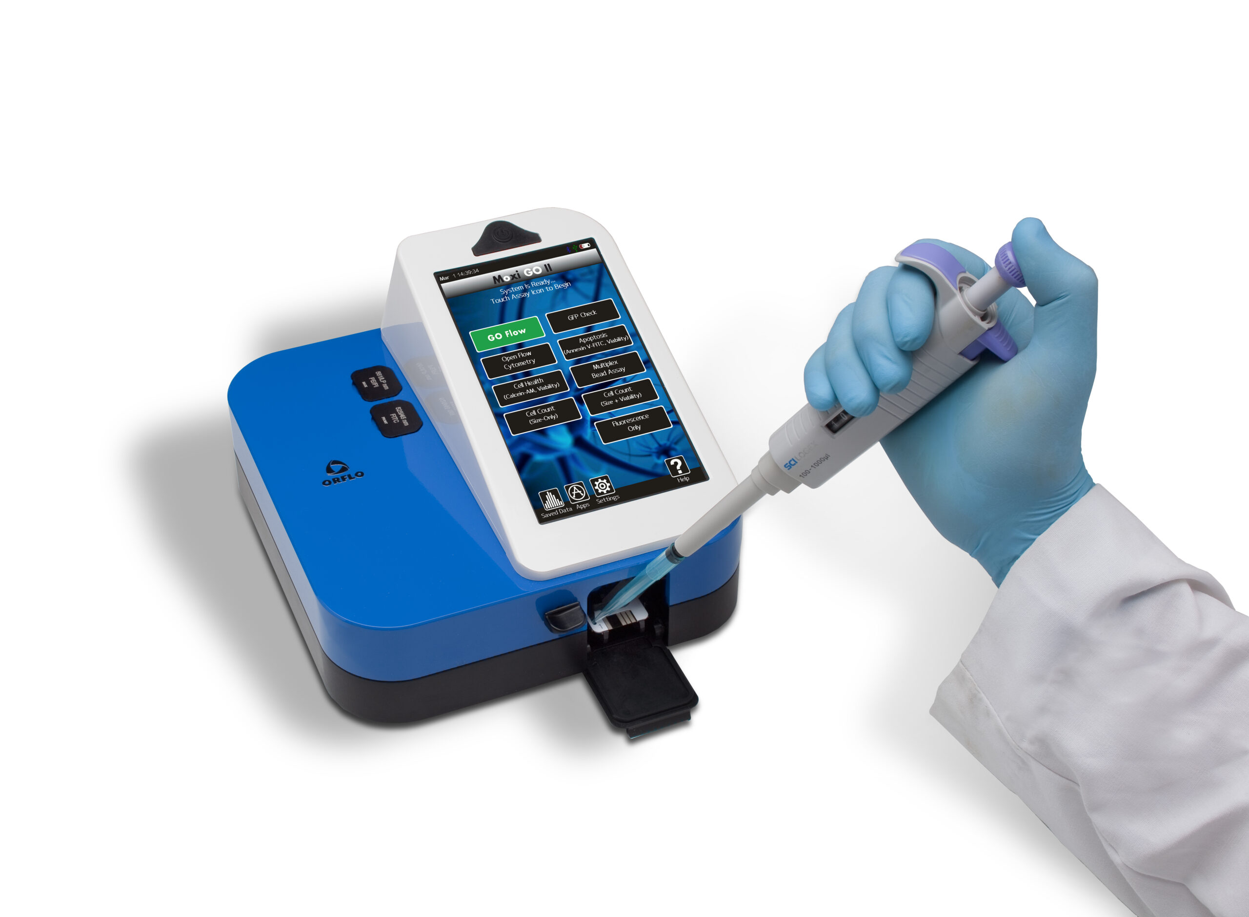
Next Generation Coulter-Principle Cell Analyzer. Combines the Coulter Principle (for highly-accurate cell counts and exact, volumetric cell sizing) with a 488nm laser and two PMT fluorescence detection channels (for cell health assays, robust CAR-T monitoring, cellular response profiling, and immunoprofiling).
The Moxi GO II’s new Auto-Gating feature will analyze results in an accurate, repeatable way to provide the most consistent results — presented in a simplified new Data Summary page. The new Batch Mode feature allows you to run multiple tests of the same sample type. It will auto-find live and dead cell populations, eliminating user-to-user variability. |
| id | MXG102 |
| Included Accessories | USB power cord, US style USB power adapter, and Type S+ cassette pack |
| AC Power Type | 110 VAC |
| Applications | Mulitplexed Bead ELISA’s|In Cell Westerrns|In Cell Protein Quant|GFP| Gold Standard Cell Count and Viability|Mito Potential|ROS|Phagocytosis |
| Battery Type | Rechargeable 3.7 V, 7500 mAh lithium ion |
| Cassette Types | Type S+ |
| Cell Particle Concentration Range | 5,000 – 1,000,000 cells/mL Type S+ |
| Cell Types Tested | HEK-293|HeLa|PC12|CD3+T|CHO-K1|Cos-7|HepG2|Hybridoma|Jurkat E6-1|K562|MCF7|Mesenchymal SC|Monocyte|Mouse ESC| NIH 3T3| PBMC (cultured)|Red Blood Cells (RBC)|L5178y| C. albicans (Yeast)| S. cerevisiae Vin 13 (Yeast)|S. cerevisiae X5 (Yeast)|Wine Yeast (natural fermentaion)|S.cerevisiae (Baker’s Yeast|Safale US-05 Yeast| |
| Data Output Formats | FCS 3.1, screen shots (.bmp), CSV |
| Data Storage Capacity | 4Gb |
| Display Resolution | 800 x 480 color touchscreen |
| Excitation Wavelengths | 488nm |
| In British Units | 10 lbs |
| Intended Use Statement | For Research Use Only. Product is not for use in diagnostic procedures |
| Laser Color | Blue |
| Measurable Dynamic Range | 3 – 27 microns Type Type S+; 4 – 35 microns Type MF-M |
| Measurement Time | 10 seconds Type S+ |
| MPI Cell Health Ratio | Yes (Size histogram only) |
| Number of Detection Channels Flow parameters | 2 color, 1 size, 1 forward extinction |
| Number of PMTs | 2 |
| Optical Detection Range | 525/45nm (e.g. FITC, GFP) and 561nm/LP (e.g. PE, RFP) |
| Particle Size Detection Method | Impedimetric (Coulter Principle) |
| Platform | Open platform: 561nm/LP (PI, PE, DS Red, Sytox Orange, 7 AAD, Nile Red, Rhodamine Red, Sun Coast Yellow, PE/Cy5), 525/45nm (FITC, GFP, Alexa Fluor 488nm, Calcein) |
| Pre-Programmed Tests | Mulitplexed Bead ELISA’s|In Cell Westerrns|In Cell Protein Quant|RFP|Gold Standard Cell Count and Viability|Mito Potential|ROS|Phagocytosis |
| Resolution Histogram Bins | 1000 |
| Sample Type | Beads|Cell Preparations |
| Sample Volume | 60 µL |
| Supported Connectivity | USB on-the-go |
| Useable Cell Volume | 14 – 10,306 fL Type S+; 14 – 22,449 fL Type MF-M |
| Weight | 4.53 kg |
How It Works
The operating principle behind the Moxi GO II Cell Analyzers is a unique combination of Coulter-style cell size determination with simultaneous fluorescence detection. As cells flow single file through the microfabricated single-use flow cell the volume of each particle is measured at the exact same time as their primary fluorescence is measured using a 488nm (MXG102) solid state diode laser with and with the following emission filters – 525/45nm (e.g. FITC, GFP, Alexa 488) and 561nm/LP (e.g. PE, RFP). Thousands of cells are measured in the 10 second read time and the data are plotted in a gradient density scatter plot as Cell size (volume) vs. Fluorescence (PMT voltage). Gating is easily performed on the unit using a interactive touch display, and the resulting live/dead ratios are automatically calculated (depending on the app selected). The analyzed data can also be displayed as a two color size histogram. Total volumetric cell counts are automatically determined for each test by precisely measuring the volume of fluid being analyzed.
Step 1:
Select desired app, insert the cassette and close the doors.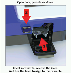
Step 2:
Once auto-alignment is complete, open the top door and pipette 60μl of labelled sample into the cassette.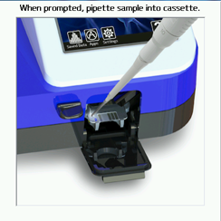
Step 3:
Close the top door, assays run automatically and results are generated in ~10 seconds. Note: Each cassette holds 2 tests. When Sample 1 is complete, simply re-insert other end of cassette into Moxi GO, and load Sample 2.
Data
Data can be displayed on the unit in both a color density scatter plot and a two color size histogram. Simply drag gates using the intuitive touch display for instant live/dead ratio calculations and each of the gated volumetric cell counts (i.e., total population, live population, and dead population (Viability App). The mean cell volume for the gated populations is also automatically displayed on the unit. Results from each test are stored in the standard FCS 3:1 format and can be viewed using any Cell Analysis analysis package. The actual Moxi Flow screenshots from each assay (dot plots and histograms) are also stored in bitmap format for use online. Hundreds of files can be stored on each Moxi GO and are easily transferred to a Mac or PC using USB on-the-go. No aditional software is required.
Moxi GO II Basic Operation | Running a Test on the Moxi GO II |
|---|---|
Home Screen Overview | Toggling Between the different |
|---|---|
Setting the Noise Gate | PBMC Cell Count and Viability |
|---|---|
Running a Viability Test – HEK’s | Setting Gates: T-Cell Example |
|---|---|
Monocyte Isolation Purity Check |
|---|